Babies Circumsized Penis Tip Ks Purple and Blacm
This commodity has been cited past other articles in ScienceCentral.
Abstruse
Kaposi's sarcoma (KS) is a multifocal hemorrhagic sarcoma that occurs primarily on the extremities. KS limited to the penis is rare and a well-recognized manifestation of acquired immune deficiency syndrome (AIDS). However, KS confined to the penis is extraordinary in human being immunodeficiency virus (HIV)-negative patients. We present the case of a 68-twelvemonth-one-time homo with a nighttime carmine ulcerated nodule on the penile skin, which was reported equally a nodular stage of KS. We detected no prove of immunosuppression or AIDS or systemic involvements in further evaluations. In his by medical history, the patient had undergone 3 transurethral resections of bladder tumors due to urothelial cell carcinoma since 2000 and total gastrectomy, splenectomy, and adjuvant fluorouracil/cisplatin chemotherapy for seven months due to avant-garde gastric carcinoma in 2005. The patient was circumcised and has had no recurrence for 2 years.
Kaposi's sarcoma (KS) was introduced by Moritz Kaposi to describe a malignant neoplasm of the vascular endothelium of multifocal origin that occurs primarily on the extremities. Earlier the acquired immune deficiency syndrome (AIDS) era, KS was a rare sarcoma that was mainly seen in Mediterranean men. In the early 1980s after the onset of the AIDS epidemic, the incidence of KS increased dramatically throughout the world [i]. Primary presentation on the penis is rare and it is more frequently observed in AIDS patients, who usually develop an aggressive form and in whom approximately ii-three% of cases bear witness penile KS lesions as the starting time manifestation of the disease [ii]. However, KS limited to the penis is extremely rare in human immunodeficiency virus (HIV)-negative patients. We experienced a instance of ulcerated nodular KS bars to the penis that was very rare in Korea. Hither, nosotros written report our example with a review of the literature.
Case REPORT
A 68-year-old man with no history of homosexual activity presented with a painless ulcerated night cherry nodule on the penile shaft noticed 3 months earlier. He had both legs amputated considering of a flop explosion during the Korean War. The patient had undergone iii transurethral resections of bladder tumor (TURBTs) due to urothelial jail cell carcinoma since he visited us with a chief complaint of painless gross hematuria in September 2000. Likewise, the patient underwent total gastrectomy with splenectomy on account of advanced gastric carcinoma in May 2005 and six treatments of adjuvant fluorouracil (5-FU)/cisplatin chemotherapy from July 2005 to January 2006. On the physical exam, an approximately ane cm sized nodule with pus-like discharge was axiomatic on the outer layer of the dorsal prepuce and two black discolorations on the glans were obvious. There was no evidence of inguinal lymphadenopathy. The results of routine laboratory investigations and urine examinations were normal. Circumcision including the ulcerated nodule was performed. Purple discoloration and profuse haemorrhage were evident on the field excised nodule. Past histologic examination, the nodule with ulceration was diagnosed as the nodular phase of KS. Histologic findings showed an infiltration composed of spindle cells scattered between collagen bundles and small vascular proliferation. Slit-like spaces containing red cells with marked nuclear pleomorphism and mitoses were observed. Immunohistochemical investigations revealed a vascular tumor considering of reasonable positivity for vascular markers such as CD31 and CD34, and immunohistochemical staining confirmed KS by diffuse nuclear staining of man herpesvirus type 8 (HHV-viii) latent nuclear antigen one (LNA-1) (Fig. i).
At the repeat concrete examination, nosotros establish no other pare lesions. We performed boosted local excision of two black discolorations on the glans. However, they were reported as keratosis. Repeated HIV tests (enzyme-linked immunosorbent assay and Western absorb) were negative. Computed tomography scanning of the chest, intestinal cavity, and pelvis failed to detect whatever visceral lesions. A 2-year follow-up did not show any illness progression or recurrence from May 2008 to June 2010.
Word
KS is a tumor of the reticuloendothelial organisation and thus information technology is vasoformative with endothelial proliferation and spindle cell germination on histologic examination. Usually, it presents as a cutaneous neovascular lesion; a raised, painful, bleeding papule; or ulcer with blue discoloration. KS is subcategorized as iv types: (1) classic KS, which occurs in patients without known immunodeficiency and has an indolent and rarely fatal class; (2) immunosuppressive treatment-related KS, which occurs in a patient receiving immunosuppressive therapy for organ transplantation or other indications and is often reversed with dosage modification of the immunosuppressive agents; (3) African KS, which occurs in young men and may be indolent or ambitious in course; and (4) epidemic or HIV-related KS, which occurs in patients with AIDS [3]. In particular, classic KS primarily affects males of Mediterranean or Eastern European descent over the historic period of 60 years [4].
In a recent study, localized classic KS of the glans penis was reported to exist rare with only 51 cases reported; 38 cases amongst them were regarded as isolated classic KS without any association with AIDS or immunosuppression [4]. However, the majority of cases of primary penile KS reported before the introduction of HIV testing are hard to classify [2]. In patients with classic KS, master penile lesions usually present as a single reddish-majestic to blueish nodule, and other clinical presentations are less common. In item, ulcerative nodules have been described in only two patients. The most often involved site is the glans, occasionally in association with swelling and lymphatic edema because of astringent involvement. Lesions may also involve the foreskin, the coronal sulcus, or the meatus. However, involvement of the shaft is rare and it is ordinarily associated with lesions located on the glans or coronal sulcus [ii]. Histologic characteristics of penile KS are similar to those of KS at other peel sites. In KS, cells that unremarkably line small blood vessels proliferate in an abnormal mode, extending outward from what would have been the lining of the vessel to penetrate betwixt and partially surround nearby collagen bundles, thus creating 'stellate' and 'ectatic' blood vessels that are not closed off just rather are open to surrounding tissue [5]. Commonly, it includes a sometimes branching, irregular, slit-like vascular space containing erythrocytes in the patch stage and prominent groups or sheets of proliferating spindle cells in the nodular stage [6].
The pathogenesis of KS is uncertain. Recent studies showed an clan betwixt all types of KS and infection of human being herpesvirus type eight (HHV-viii), known as KS-associated herpes virus (KSHV) [ane-4,7-10]. HHV-eight as a potential oncogenic virus is supported by its tight relationships with chief effusion lymphoma (PEL), its homology with ii other oncogenic gamma herpesviruses (herpesvirus saimiri and Epstein-Barr virus), and its ability to alter the growth of human being endothelial cells in vitro [7]. In add-on, AIDS-related multicentric Castleman's illness, multiple myeloma, and lymphoma are other neoplastic disorders in which HHV-8 has been usually establish [8]. Therefore, infection of HHV-viii is a pathogenetic factor in KS considering its potential equally an oncogenic virus. It seems that the route of HHV-viii manual may exist both sexual and nonsexual. High HHV-8 seroprevalence in individuals with high-risk sexual activity represents the sexual route, and the detection of HHV-8 antibodies in children without sexual action suggests the nonsexual route. Zargari reported that saliva could be a potential source of spread of HHV-8 in the general population [7].
In our example, merely a single, ulcerated dark reddish nodule at the penile shaft virtually the coronal sulcus was diagnosed equally KS. In that KS occurs on the penile shaft with ulceration, this presentation is unusual and a rare course of chief penile KS in an HIV-negative patient. Our patient had undergone treatment for urothelial cell carcinoma and gastric cancer including chemotherapy. However, he presented with KS about 3 years later his last chemotherapy handling, and his laboratory findings had been normal since his terminal chemotherapy. Therefore, the KS of our patient seemed to be the classic type. Histologic examination showed a nodule of spindle cells with intracytoplasmic lumina and many mitoses. Immunohistochemical investigations revealed positivity for CD31, CD34, and HHV-viii LNA-one, which is known as a positive marking for KS. The patient vigorously denied sex since his wife died.
No standard treatment guideline for primary penile KS is available. Treatment includes local surgical excision, radiotherapy, chemotherapy, and laser therapy. Treatment with adjuvant alpha or beta interferon has also been used in some cases. In general, surgical excision is recommended for a small solitary lesion, whereas bourgeois radiations therapy may exist useful for large lesions. Systematic chemotherapy is usually reserved for more avant-garde cases with visceral involvement or generalized lesions. The clinical course of primary penile KS is variable, and no consistent follow-up data exist. Nevertheless, local recurrences are rare if the lesion is completely removed. Onset of afar new lesions may exist observed after a period of about i-two years [two]. Our patient underwent circumcision including the ulcerated nodule and local excision of blackness discoloration on the glans. No other adjuvant therapy was performed. He has even so not presented with whatever illness progression or new lesions during a 2-year follow-upward.
In conclusion, primary penile classic KS in HIV-negative patients is rare and is associated with HHV-viii infection. It should be treated aggressively past surgical excision or laser therapy or radiation therapy or chemotherapy because of an uncommon association with systemic interest.
Figures and Tables
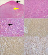
FIG. i
Histopathologic findings. (A) Ulcerated pare (black arrow) with nodule of spindle cells (xanthous arrow) (H&Due east, ×100). (B) Spindle cell proliferation with intracytoplasmic lumina containing blood-red cells (H&E, ×200). (C) Spindle jail cell proliferation with many mitoses (black pointer) (H&Eastward, ×400). (D) The neoplastic cells are positive for the vascular marker, CD31 (×200). (Due east) The neoplastic cells are positive for the vascular marking, CD34 (×200). (F) The neoplastic cells are strongly positive for man herpesvirus type viii latent nuclear antigen ane (×200).
Notes
References
1. Gönen M, Cenker A, Kiyici H, Kalkan M. Penile Kaposi's sarcomas in a circumcised and HIV-seronegative patient. Int J Urol. 2006. 13:318–320.
two. Micali G, Nasca MR, De Pasquale R, Innocenzi D. Primary classic Kaposi's sarcoma of the penis: report of a case and review. J Eur Acad Dermatol Venereol. 2003. 17:320–323.
3. Pettaway CA, Lynch DF, Davis JW. Wein AJ, Kavoussi LR, Novick AC, Partin AW, Peters CA, editors. Tumors of the penis. Campbell-Walsh urology. 2007. ninth ed. Philadelphia: Saunders;959–991.
4. Pacifico A, Piccolo D, Fargnoli MC, Peris 1000. Kaposi's sarcoma of the glans penis in an immunocompetent patient. Eur J Dermatol. 2003. thirteen:582–583.
five. Schwartz RA, Cohen JB, Watson RA, Gascón P, Ahkami RN, Ruszczak Z, et al. Penile Kaposi's sarcoma preceded past chronic penile lymphoedema. Br J Dermatol. 2000. 142:153–156.
6. Lucia MS, Miller GJ. Histopathology of malignant lesions of the penis. Urol Clin Due north Am. 1992. nineteen:227–246.
seven. Zargari O. Exclusive penile Kaposi's sarcoma: report of an HIV-negative man successfully treated with radiotherapy. J Eur Acad Dermatol Venereol. 2006. 20:318–320.
8. Chitale SV, Peat D, Meaden JD, Johnson HB, Burgess NA. Kaposi's sarcoma of the glans penis in an HIV negative patient. Int Urol Nephrol. 2002. 34:251–253.
9. Morelli L, Pusiol T, Piscioli F, Höfler H, Weirich G, Werner M, et al. Herpesvirus eight-associated penile Kaposi'south sarcoma in an HIV-negative patient: first study of a solitary lesion. Am J Dermatopathol. 2003. 25:28–31.
x. Lee KB, Lee HS, Lee HE, Park SY, Chung JH, Choe G, et al. Immunohistochemical characteristics of Kaposi sarcoma and its mimicries. Korean J Pathol. 2006. twoscore:361–367.
Source: https://synapse.koreamed.org/articles/1005572
0 Response to "Babies Circumsized Penis Tip Ks Purple and Blacm"
Post a Comment